The blood is 5 days old and completely untreated. It is not the first time I have seen these cells. But at first I thought they might be impurities. This does not seem to be the case.
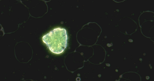
Here is a similar looking cell, in a different color.
A cell inclusion at 400x magnification
A conspicuous orange-colored inclusion that could possibly be iron.
What can be observed more and more frequently after a blood sample is that these dots begin to wrap around the white blood cells and circle them in an anticlockwise direction. - see video - This leukocyte also has a small crystal directly on the cell.
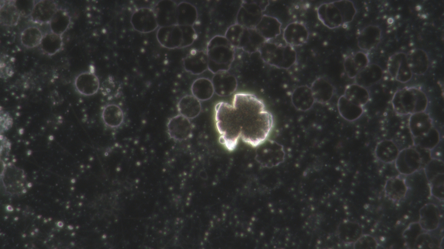
After another 3 days, this cell looked like this:
The following effect was observed by chance. If you turn down the microscope's LED lamp, the entire black structure inside begins to contract, as can be seen in the following movie.
However, this can only be repeated until the build-up process is complete. At the end, this substance seems to be transformed back into a crystalline form, so that no more movements can be dissolved.
Here is a repeat attempt with stronger exposure - you can see that the cell view no longer shows any changes. What still changes is the reaction of the plasma around it. It is important to note that the light was not changed by the setting on the microscope, but by a sheet of paper that was pushed over the lower light exposure. It can therefore not have been caused by a direct influence on the microscope itself.
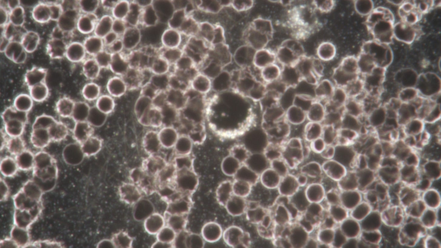
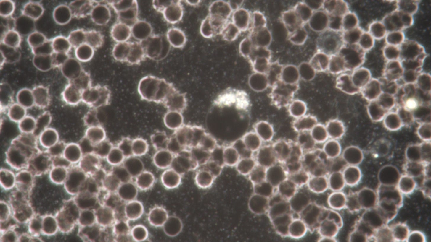
Otherwise, most white blood cells have been converted to the following state:
A giant structure surrounded by leukocytes.
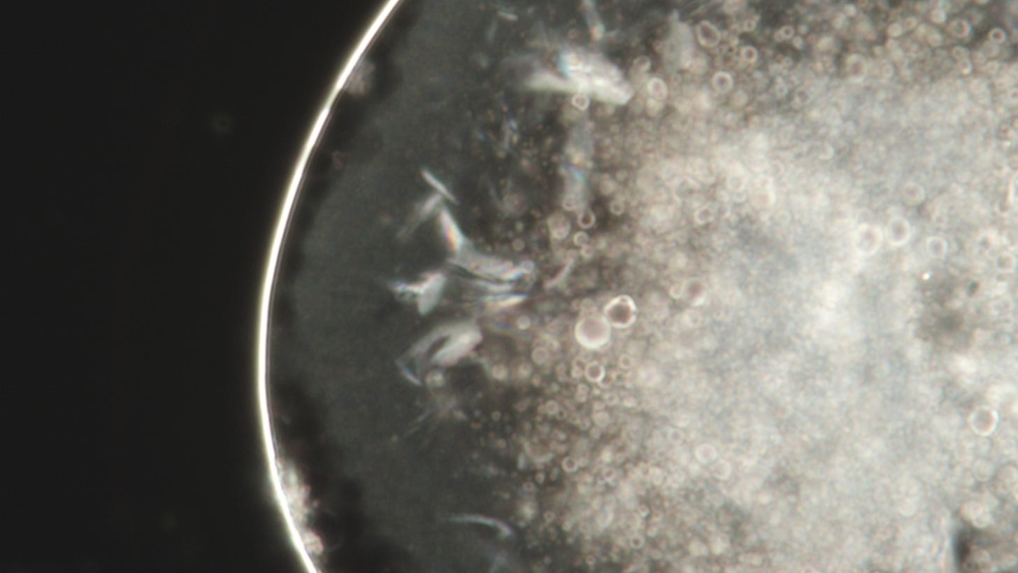
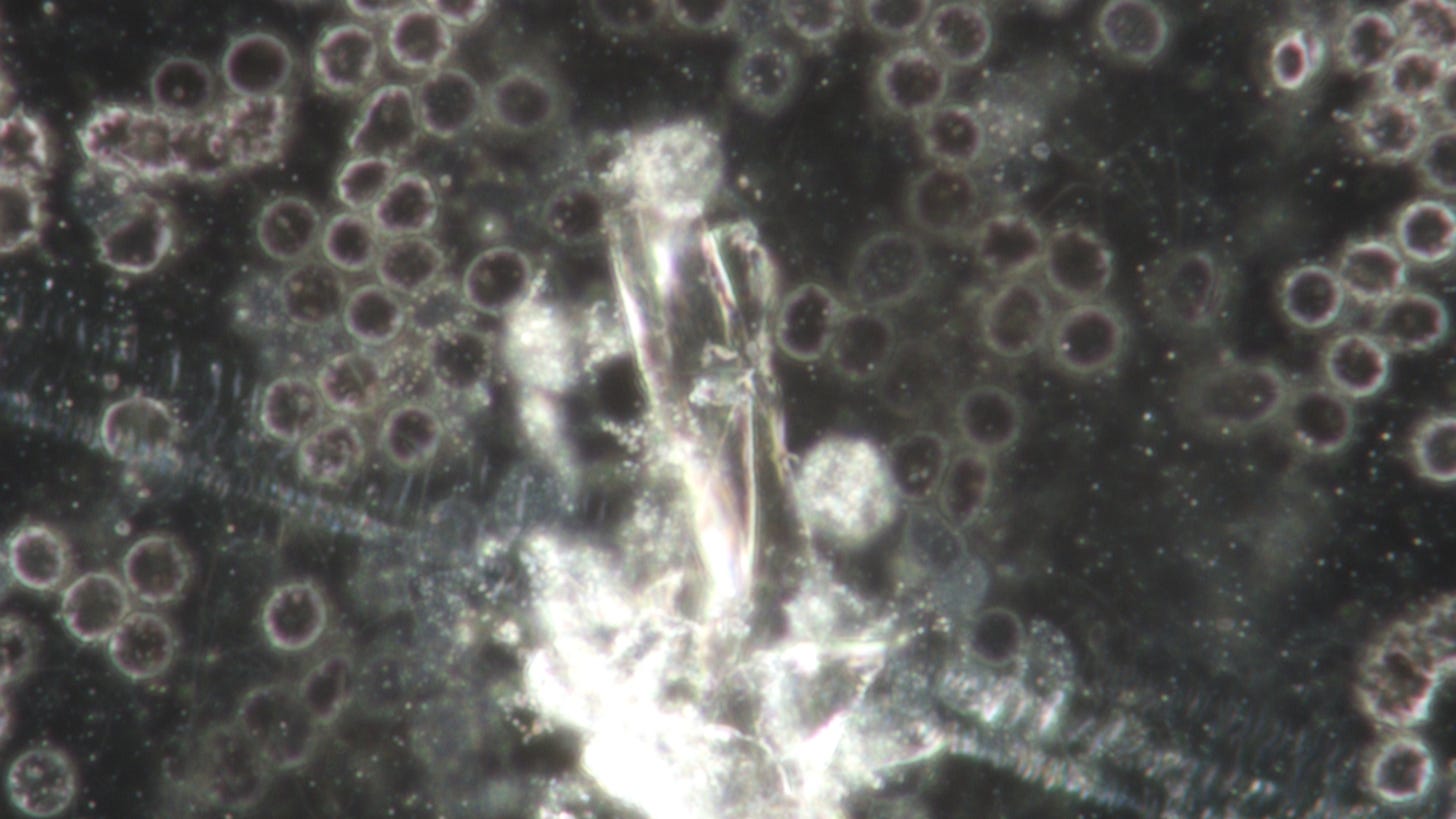
Finally, a crystal image:
The longer you look at these blood pictures, the more questions arise.





beautiful presentation and work. Thank you!