Attempt at an interpretation
In the monochrome staining with methylene blue, all protein-containing structures appear dark blue. The cell nuclei are strongly stained, the cytoplasm only weakly.
At the molecular level, methylene blue binds to negatively charged molecules, especially phosphates in the DNA. This enables detailed visualization of cell nuclei and the detection of microorganisms
The cytoplasm generally does not take on a strong color.
Methylene blue stains basic (alkaline) cell components, such as the DNA in cell nuclei, blue.
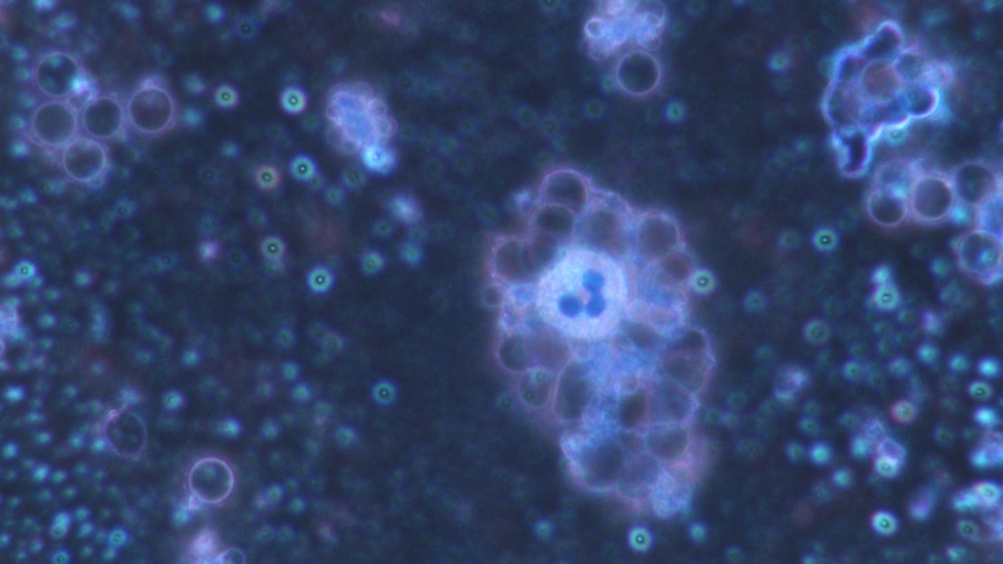
Above a video where you can see a normal leukocyte at work and to the right a cell with a brightly colored nucleus - but with a round red dot pattern.
This could correspond to this cell, which would then correspond to a remodeled leukocyte:
They all stand still - and are round as a ball, which is a sign that they are not working.
Eosin is the dye that binds to acidic cell components, such as the cytoplasm of cells, and stains them red. However, there is no extra eosin in the methylene blue I use - although it is present in this compound.
Heavily stained structure in the blood - which are usually seen as hydrogels...
However, strong red stains appear in the blood - which either indicates that eosin is present there, or:
Or the following may have happened:
In an alcoholic solution (e.g. from fermentation processes) of methylene blue in an alkaline reaction, the red reduction stage of methylene blue is formed, It is assumed that this red state form of methylene blue corresponds to the first reduction stage of the methylene blue cation to leucomethylene blue the methylene blue molecule.
In any case, some extremely red colorations are clearly visible.
On the other hand, there are now polymer-based nanocapsules for passive drug delivery that work with eosin. These areas would then also light up red. The converted leukocytes in particular should be considered here - they now have red inclusions in the cell area.
In the video above, the staining clearly shows how an artificial film covers parts of the blood picture that are in contact with a bundled strand.
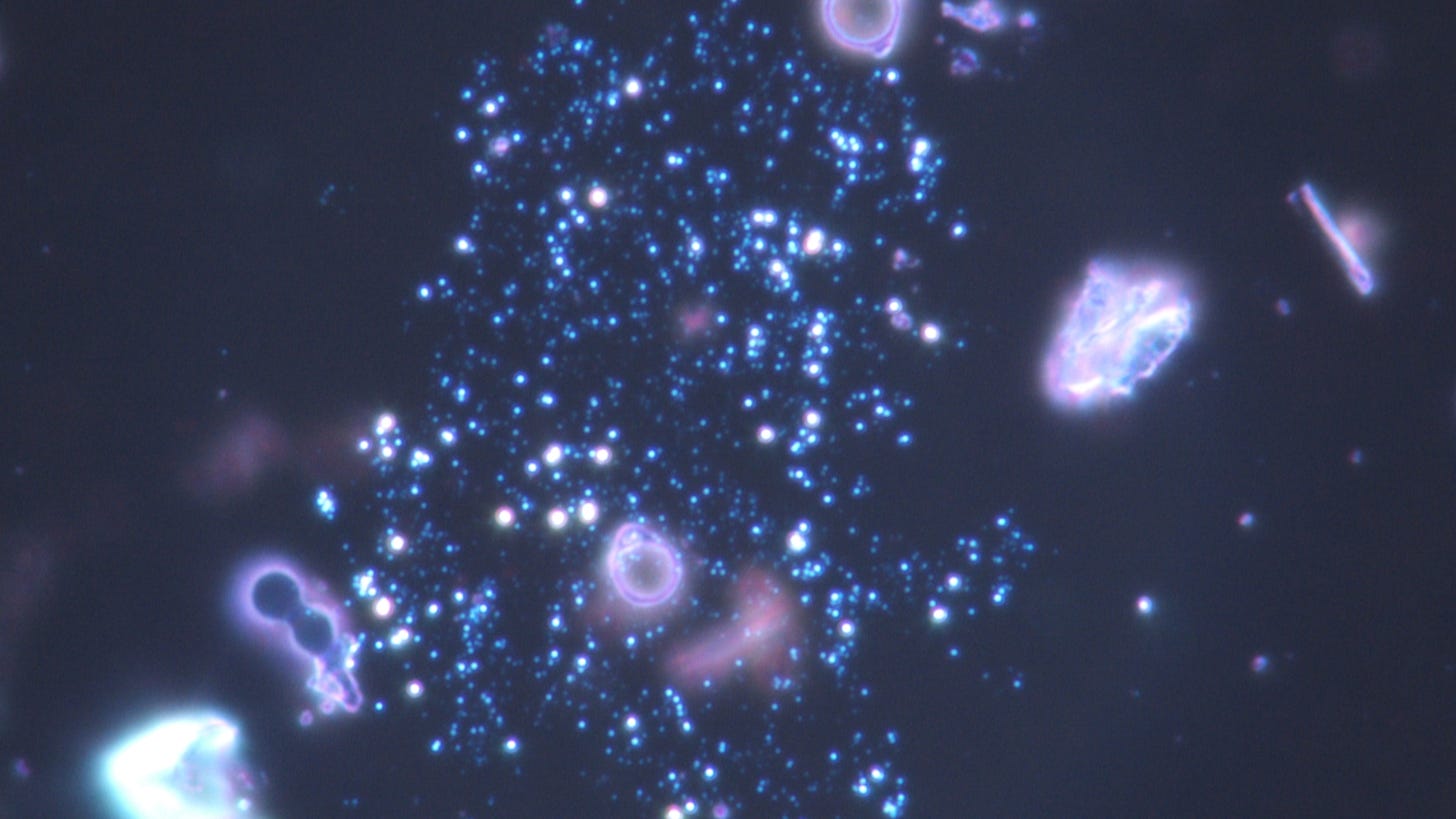
Below you can see a structure with a bubble full of quantum dots cavorting in it.
The chemical reaction - of whatever - can be seen here at the edge of the specimen, which otherwise only occurs within the bubble formations.
This bubble formation shows a good intact membrane - otherwise the methylene blue would have reached the inside and would have colored the inner part as well.
And a few more photos and recordings from the rehearsal:
Sometimes you really want to be able to look at normal blood again...


Hi Sylvia!
Great work and analysis! Nice, but so frightening pictures and videos!
First video:
The eosin stained cell gives us a glimpse of what’s inside of these otherwise over exposed cells’ rings. Congratulations! Very good job!
This bubble formation shows a good intact membrane… :
Were the bubbles not stained because they are somehow protected by the surrounding “black pool” material (lipid?), its edge seemingly having absorbed the methylene blue to some extent?
Greetings!
Great work & good clarity ..
methylene blue has many other properties & I believe beneficial for donating electrons in the body .
Thank you for your investigatory knowledge shared with us all ..