The anatomy of the willow plant as a model for hybrid filaments in the blood?
Coincidences? They don't exist...

Anatomical similarities have led me to new questions.
Because of Eric's last article (https://substack.com/@firemedic8/p-163159128 - good job by the way!) , I went out and got one of the first sprouting leaves of Salix herbacea (herbaceous willow) and put it under the microscope to observe it more closely.
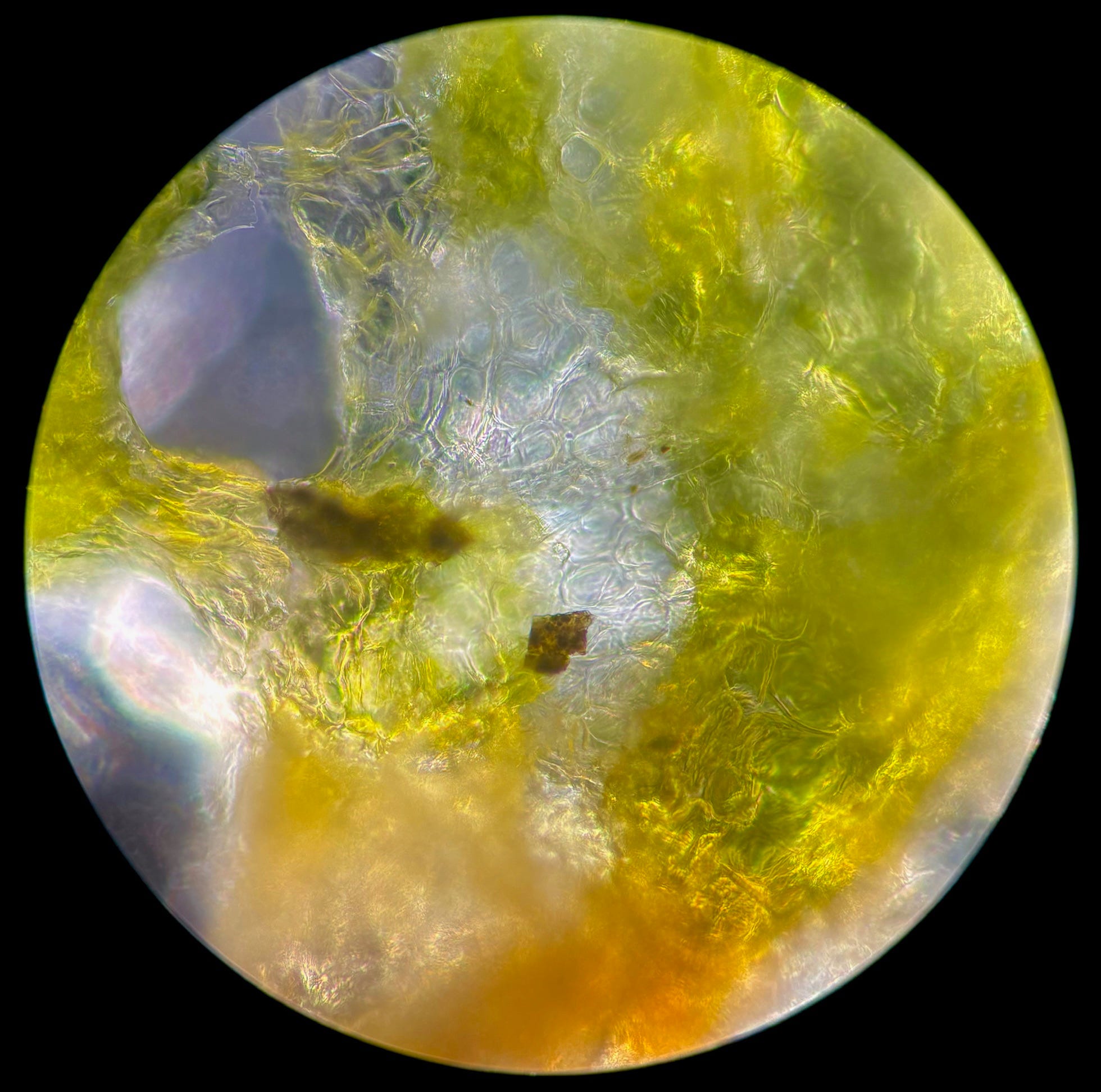
Here is a first glimpse of one of the preparations:

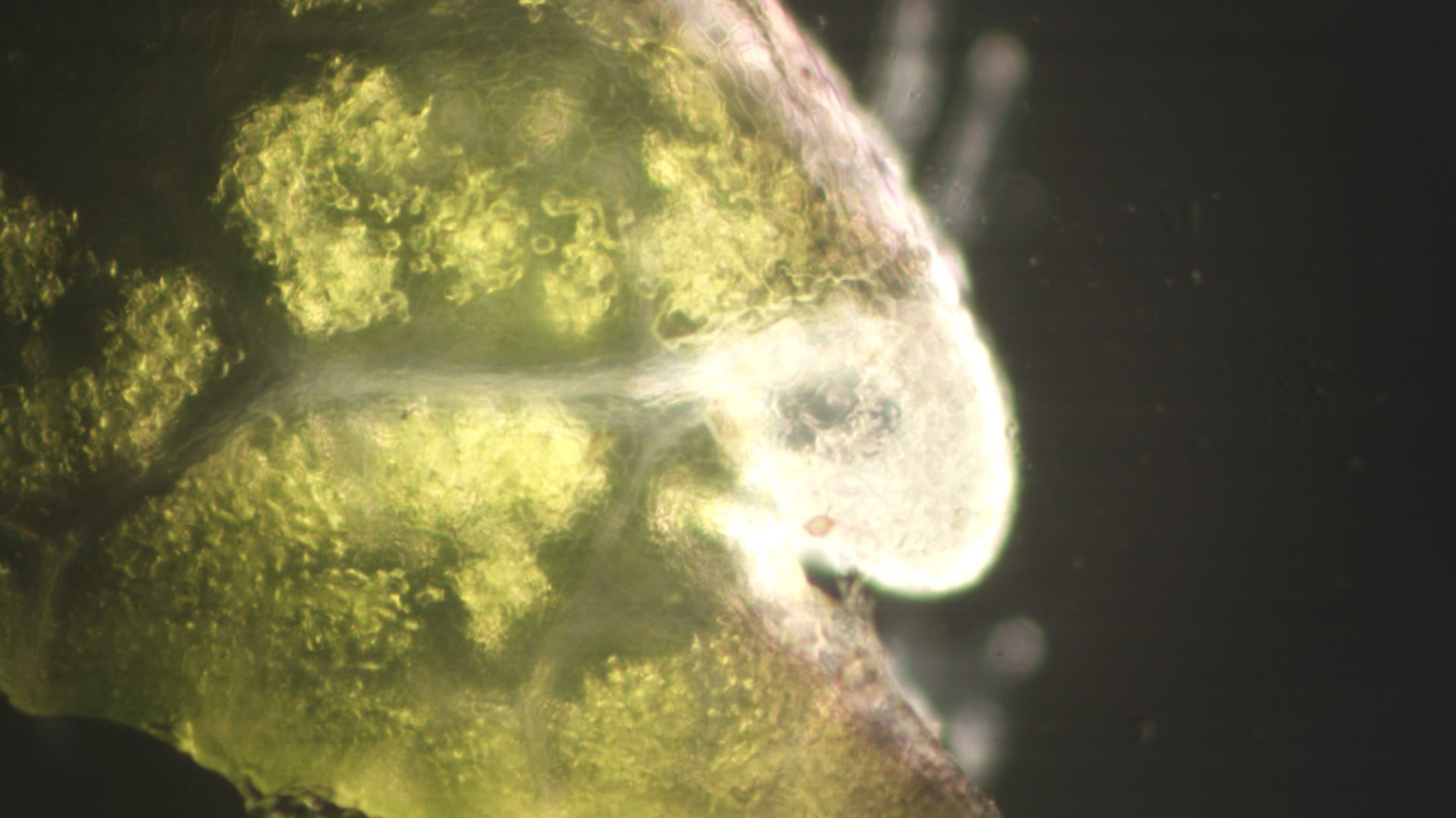
In the picture you can see something at the lower edge that resembles a mushroom-like growth:
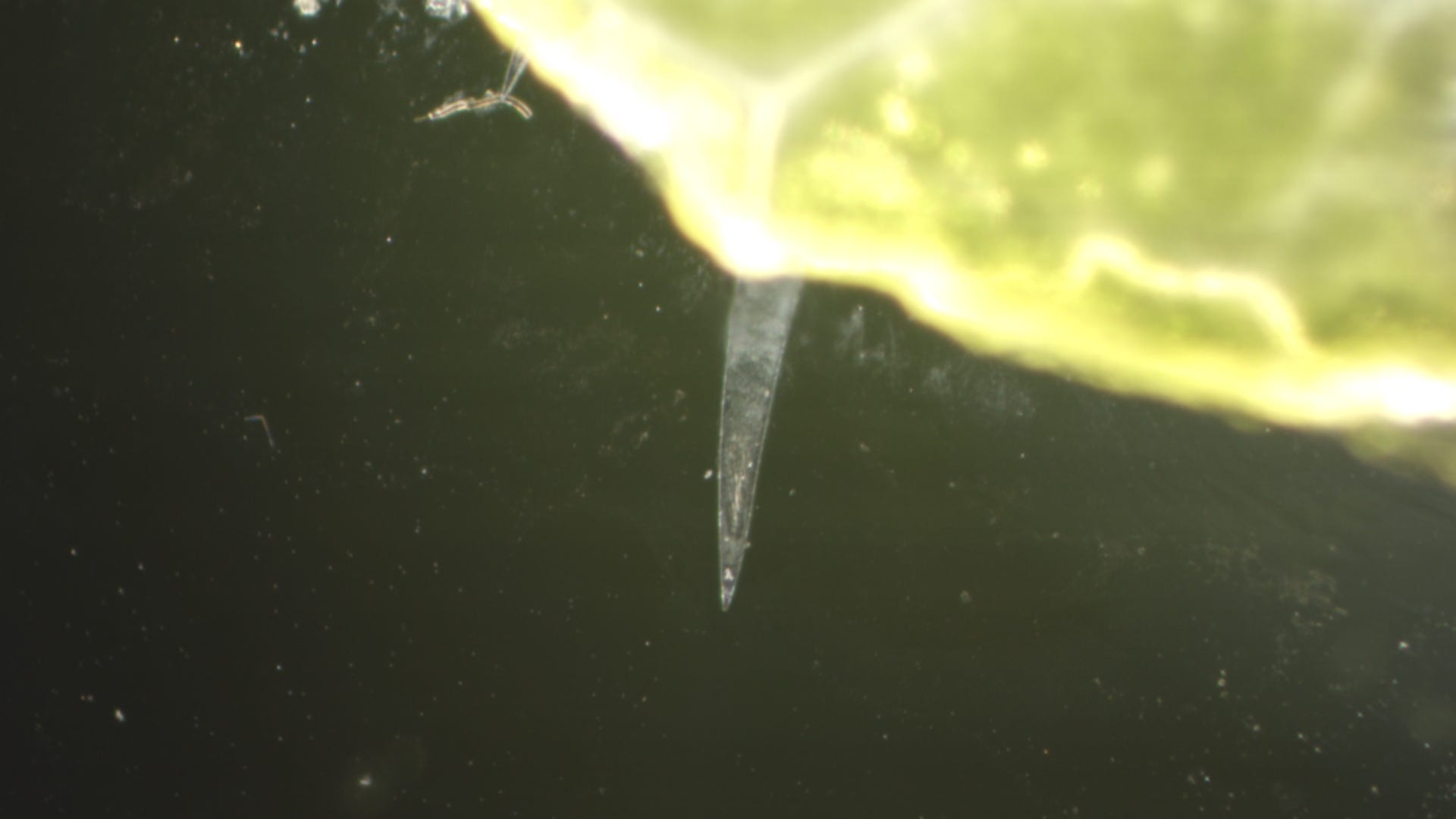
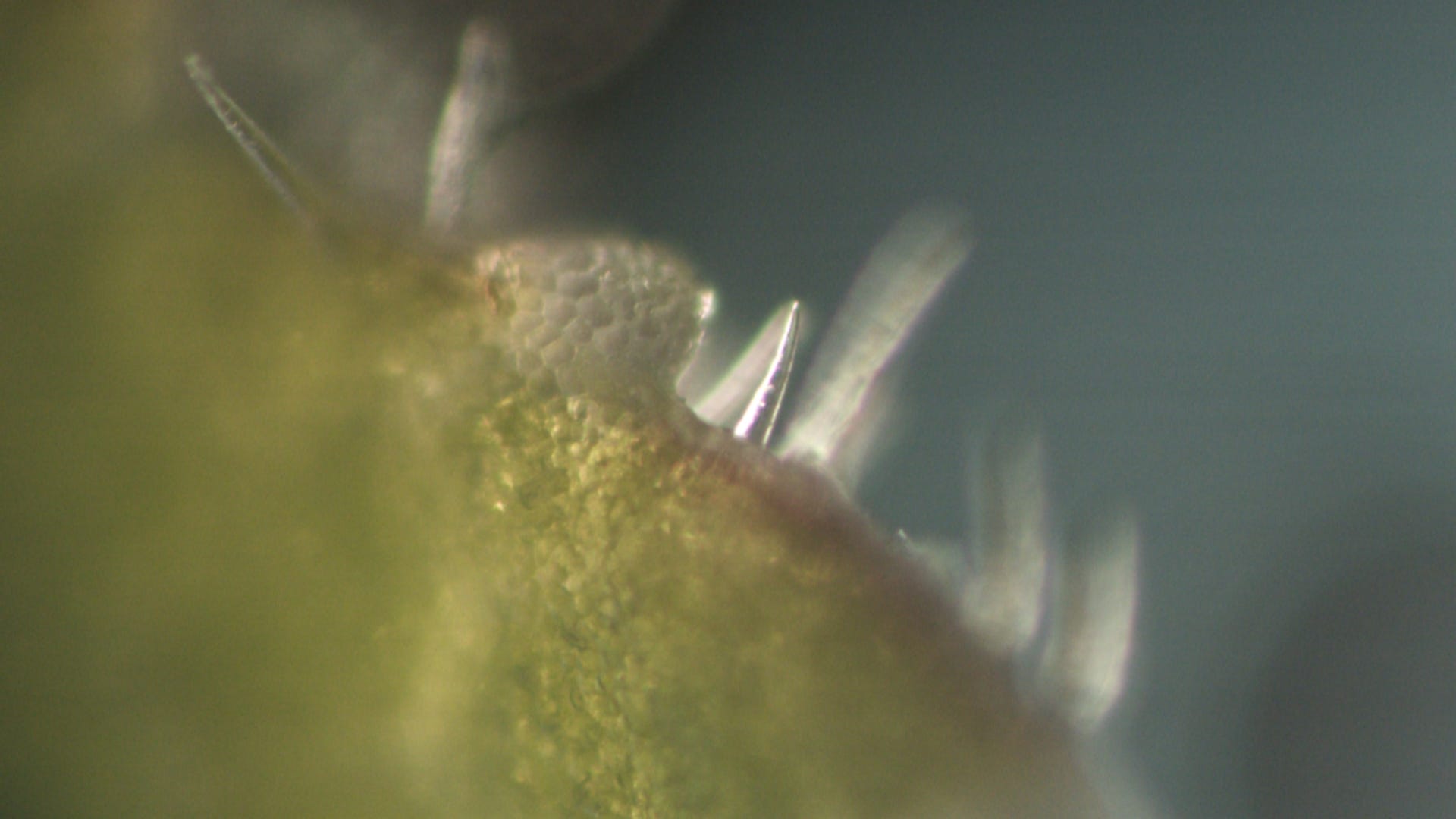
The following picture shows the small hair-like protrusions that can be seen everywhere:

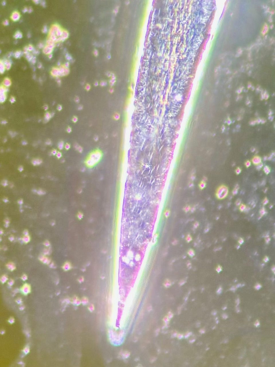
The enlarged image above shows small structures in the tip area, magnified 100x - oil objective:
This is a highly magnified video of the tip with the self-organizing particles:
These tips or small hairs that are spread over the whole leaf have small hairs that I first assumed were pores, but then I found some connections and similarities that I had already found in the blood:
I had already seen exactly this arrangement in the blood months ago. This is a thread structure from the blood at a magnification of 100x:
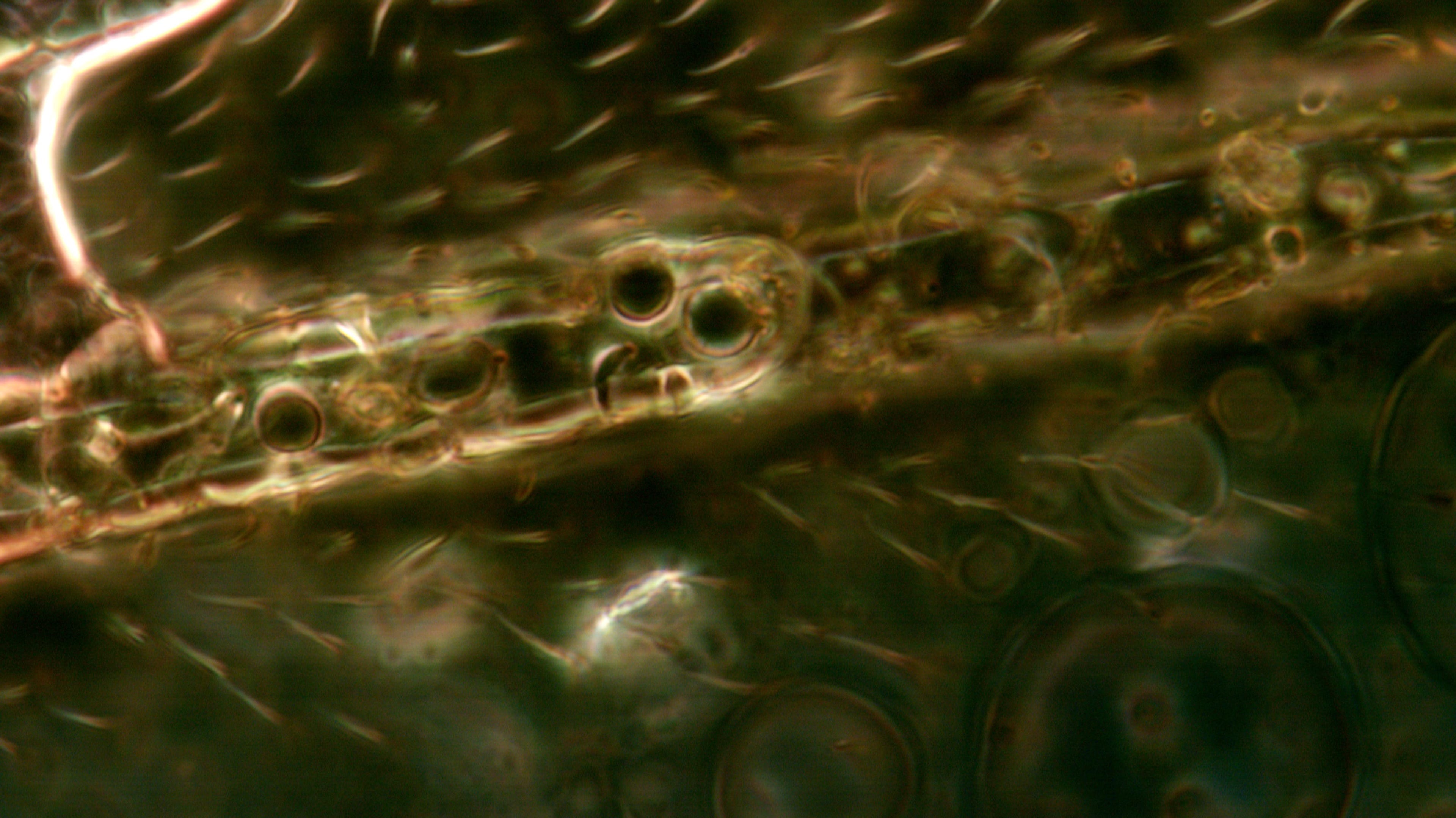
And here again for comparison in the plant:
This made me wonder whether there was a connection between the plant and nanotechnology. And I found what I was looking for:
A direct link between nanotechnology and the plant Salix herbacea (dwarf willow) has not yet been established or documented in the scientific literature. However, there are links with other willows, such as studies on the green synthesis of nanoparticles with Salix alba.
1. biogenic nanoparticle production
Some plant species are used for the “green synthesis” of nanoparticles - this means that plant extracts serve as environmentally friendly reducing agents for the production of e.g. silver or gold nanoparticles.
The genus Salix (willows) contains many species that are rich in polyphenols and flavonoids.
2. Biological inspiration in nanotechnology
Salix herbacea is one of the smallest woody plants and has an extreme adaptability to alpine/extreme environments. The microstructures of its leaves or cell walls could hypothetically serve as a model for biomimetic materials in nanotechnology.
3. Pharmacological applications
Some Salix species contain salicylates, precursors of aspirin. If attempts were made to integrate active substances from Salix herbacea into nanoscale carrier systems (e.g. liposomes or nanoparticles), there could be an indirect link to nanomedicine.

Several scientific studies have dealt with the production of nanoparticles using Salix alba:
Silver nanoparticles (AgNPs): One study reported the production of silver nanoparticles from the bark of Salix alba. These nanoparticles showed an average size of 29-35 nm and possessed antimicrobial properties against bacteria isolated from dental plaque.
They were synthesized by adding silver nitrate solution to an extract of willow bark with constant stirring and heating.
https://www.orientjchem.org/vol32no3/green-synthesis-of-silver-nanoparticles-using-bark-extract-of-salix-alba-and-its-antimicrobial-effect-against-bacteria-isolated-from-dental-plaque/?utm_source=chatgpt.comGold nanoparticles (AuNPs): Another study reported the preparation of gold nanoparticles by the reaction of gold salts with an extract from the leaves of Salix alba. The resulting nanoparticles were 50-80 nm in size and possessed antimicrobial and anti-inflammatory properties.
Green Synthesis of Silver Nanoparticles using Bark Extract of Salix Alba and Its Antimicrobial Effect Against Bacteria Isolated from Dental Plaque
Green synthesis and biological activities of gold nanoparticles functionalized with Salix alba https://www.sciencedirect.com/science/article/pii/S1878535215001975?utm_source=chatgpt.com
Taken together, it can be said on the basis of pure observations that there is a connection here and that there is a genetic component in the hybrid filaments in the blood, perhaps also in those found in the intestine, of plants such as willows.
Here are a few more pictures from the preparation:
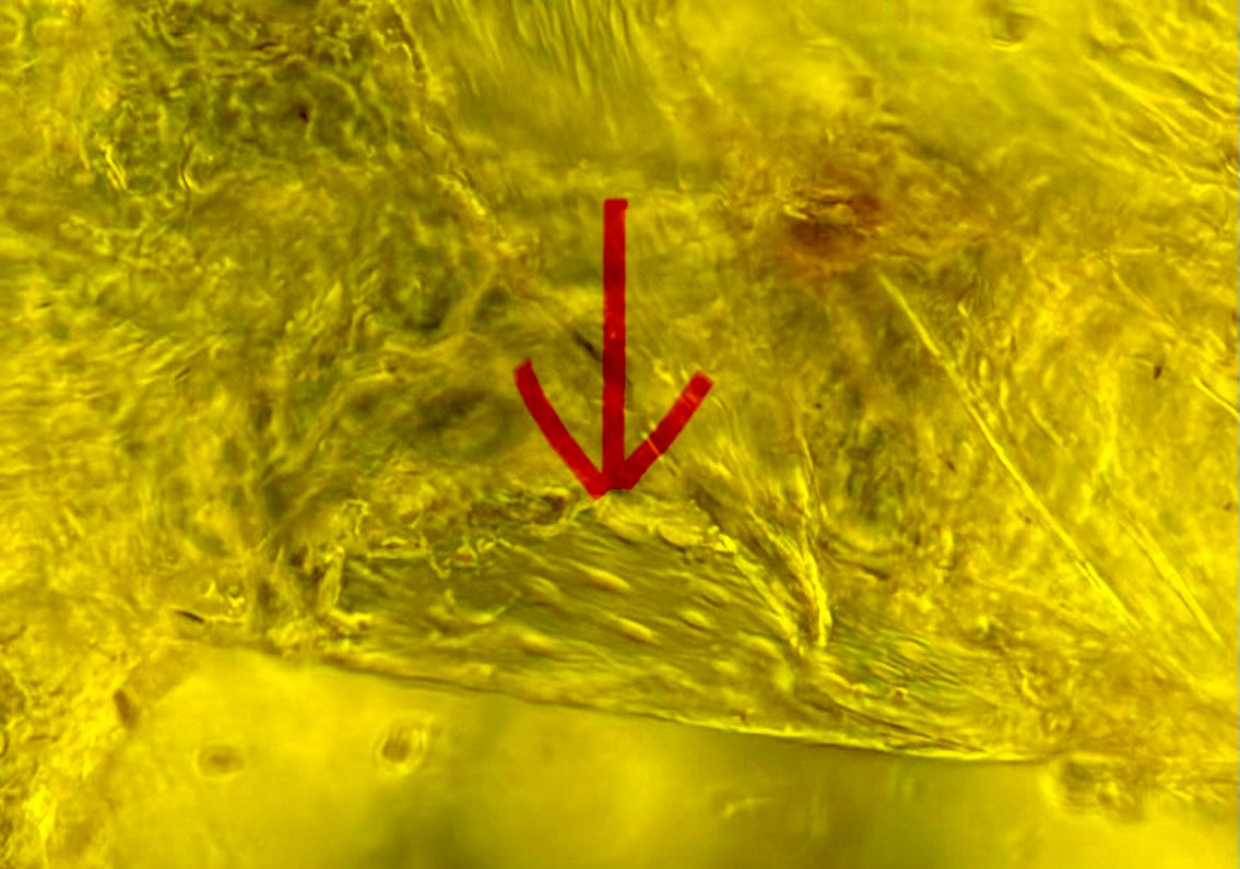
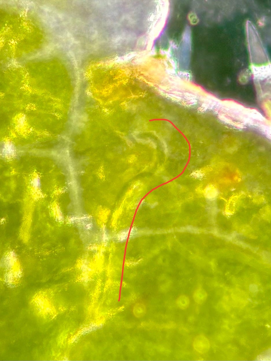
In the following picture, a blue thread was clearly visible, which I traced with the red line. It simply disappeared after a while. Where did it go? I have no idea...
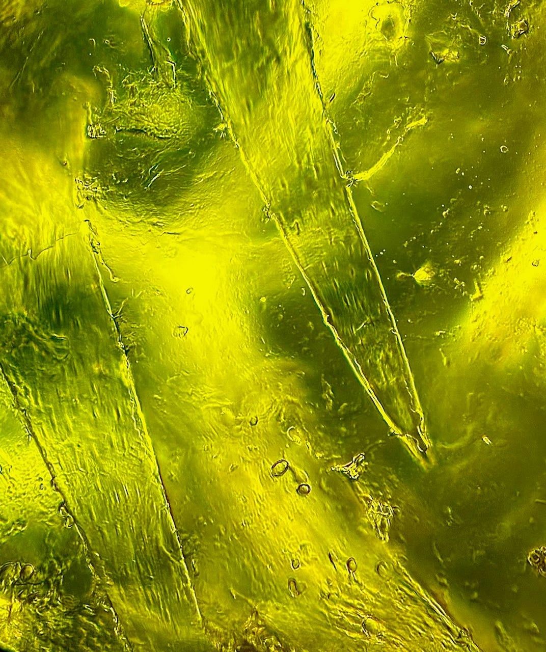
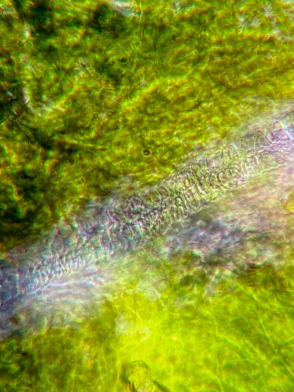
This is a close-up of a supply channel within the leaf structure. Enlarged 100 times and also zoomed in digitally:
For your help and support here on Substack you will also get access to my Future Targets, Extra Targets and the first blueprint for the LaserCube on my website.
I have set up a Telegram channel - where I post new images from darkfield microscopy. If you would like to browse through new pictures in between, you are welcome to register.