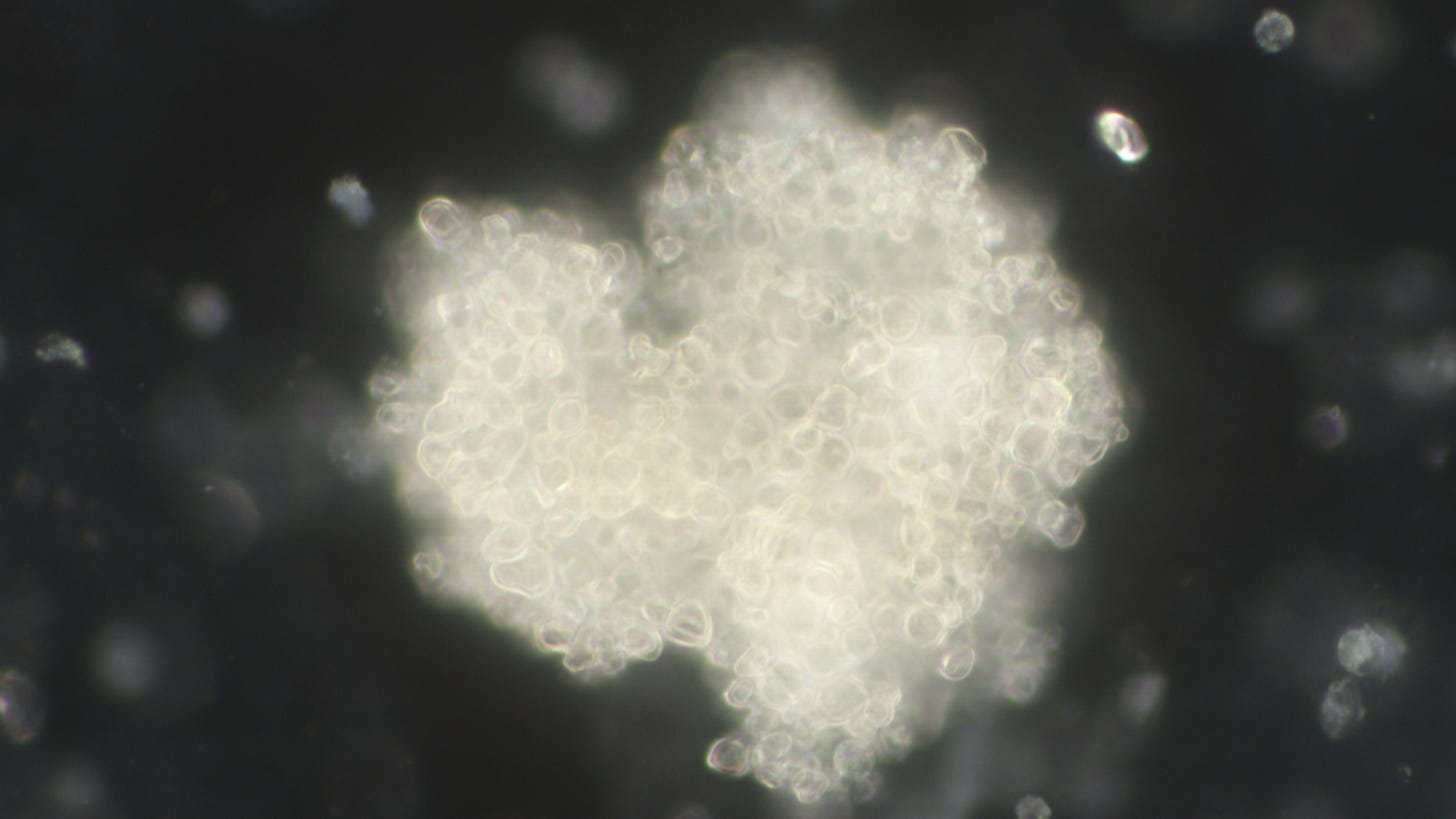
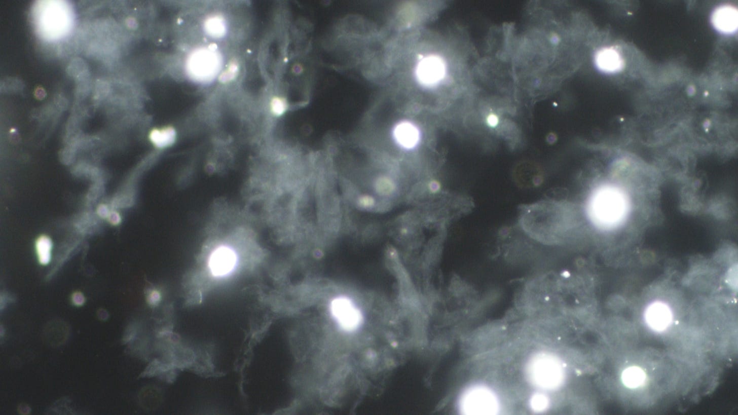
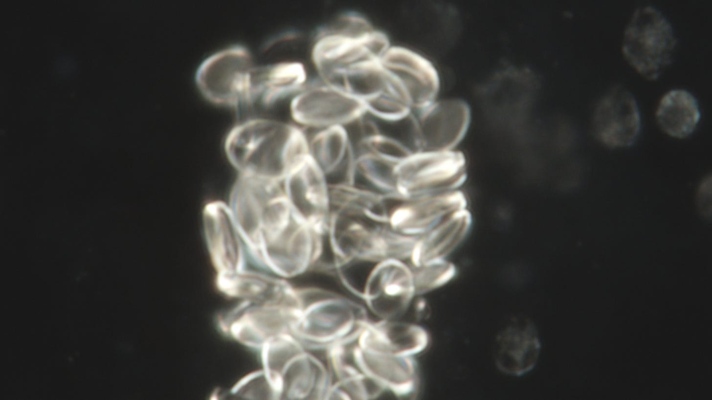
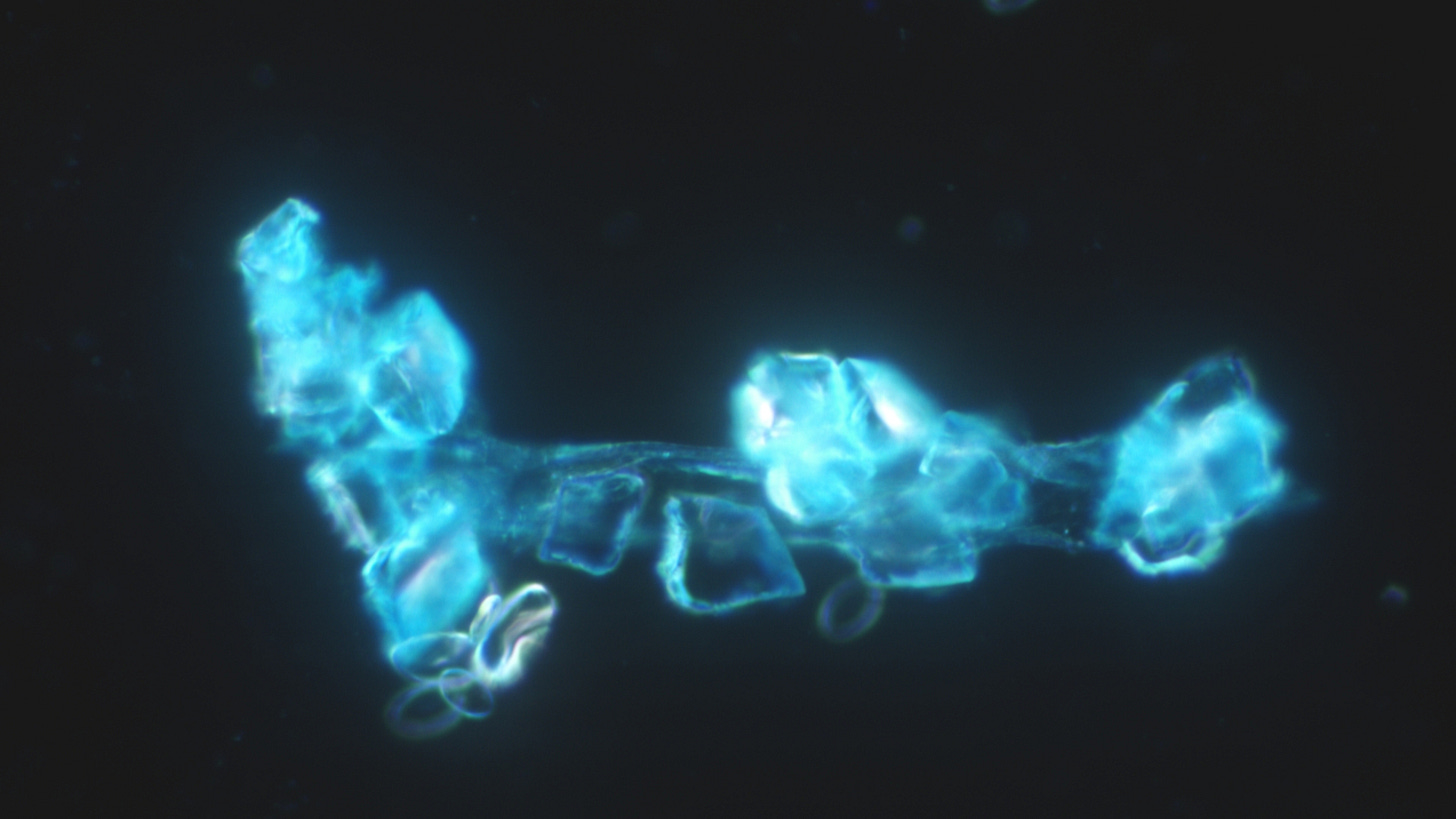
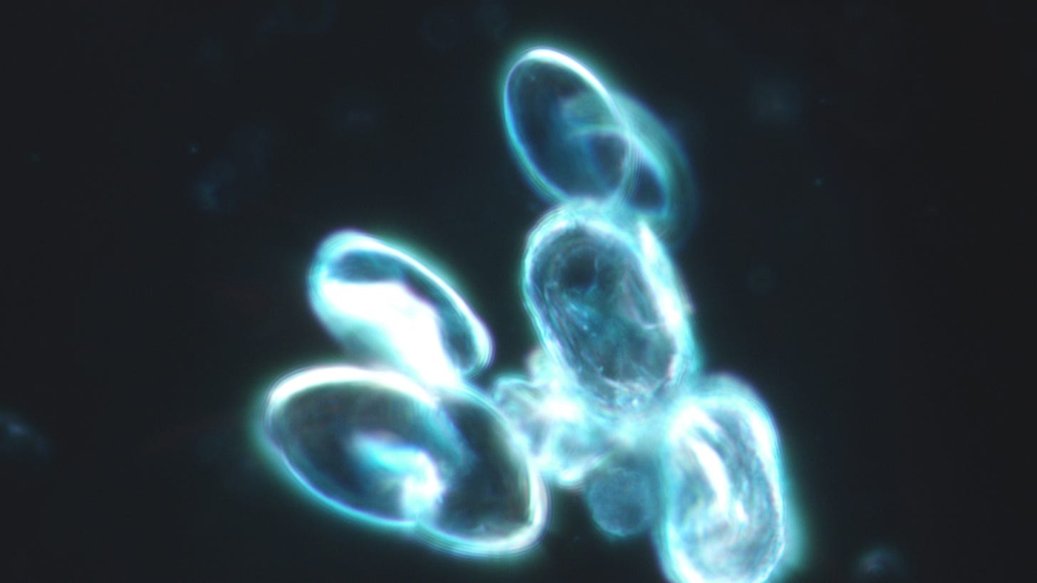
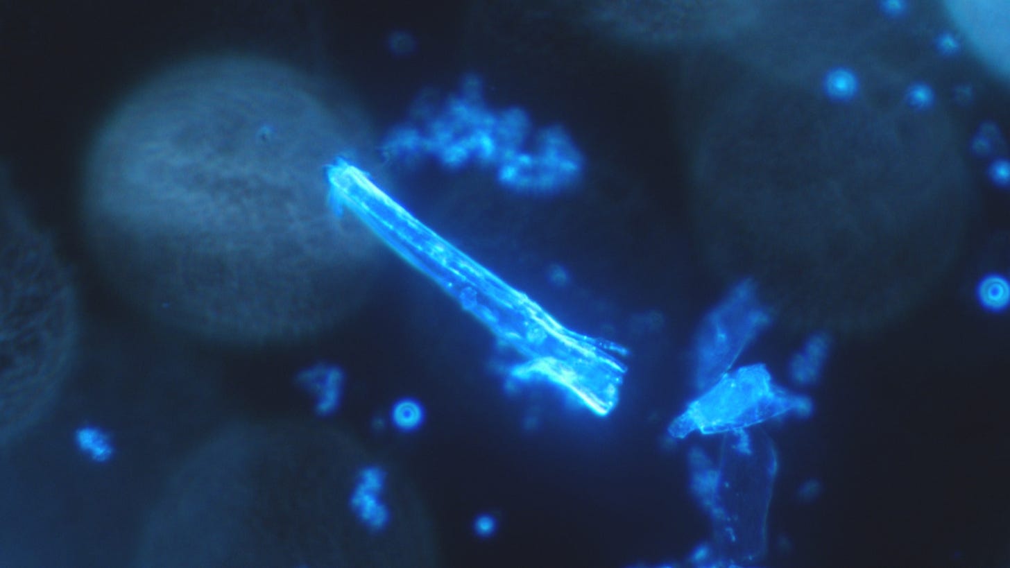
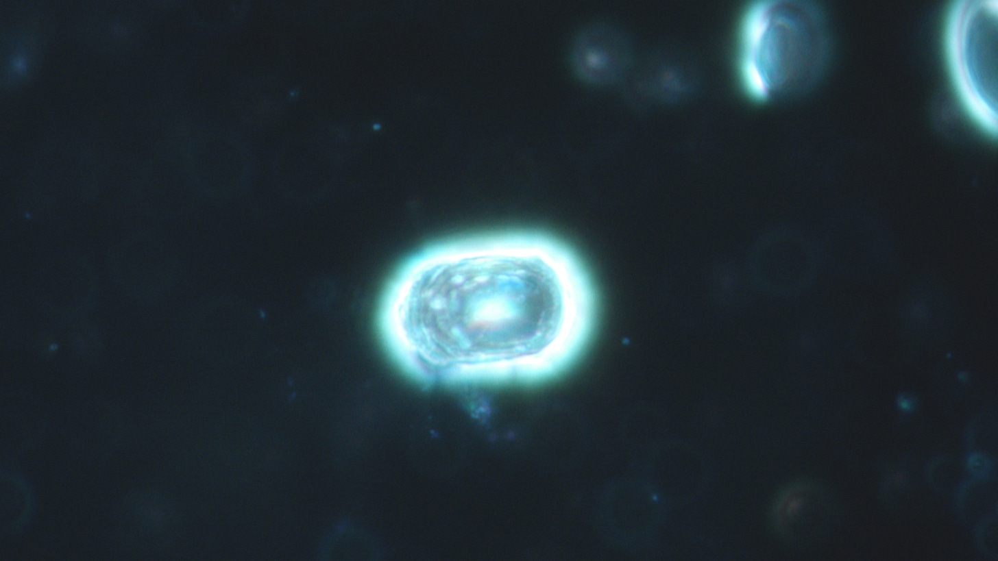
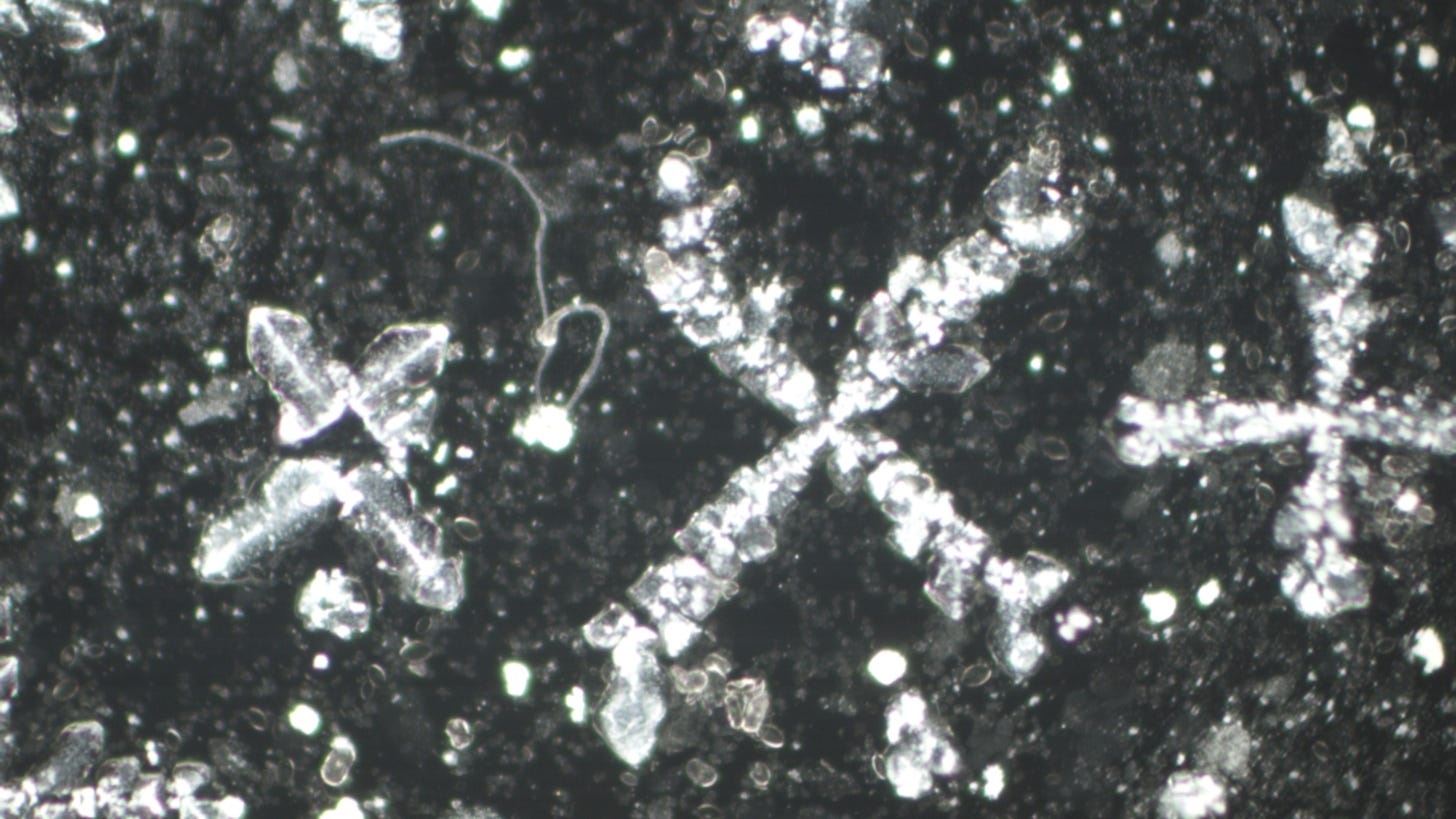
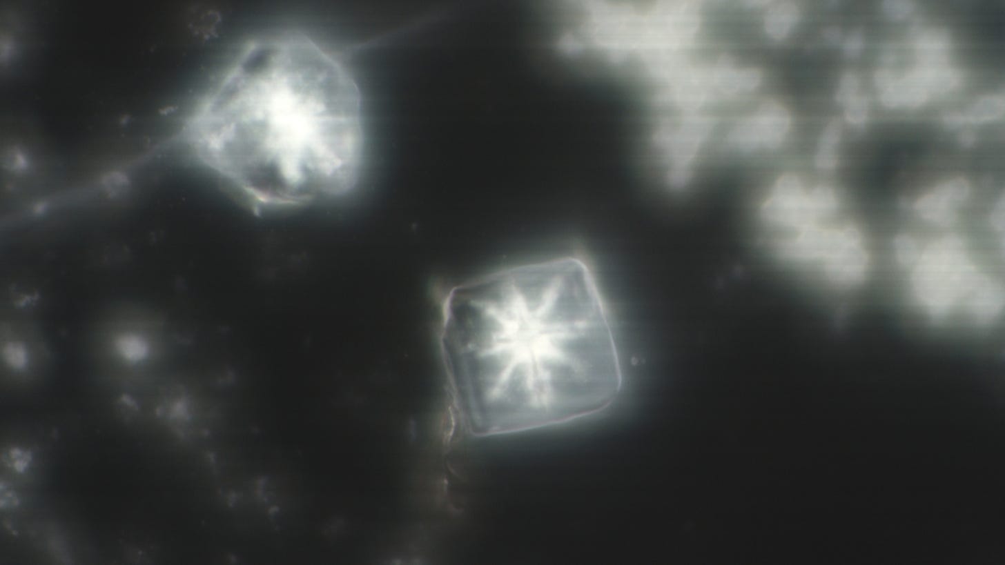
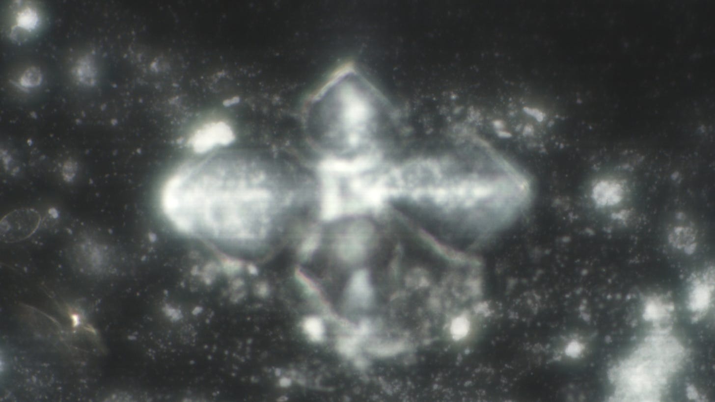
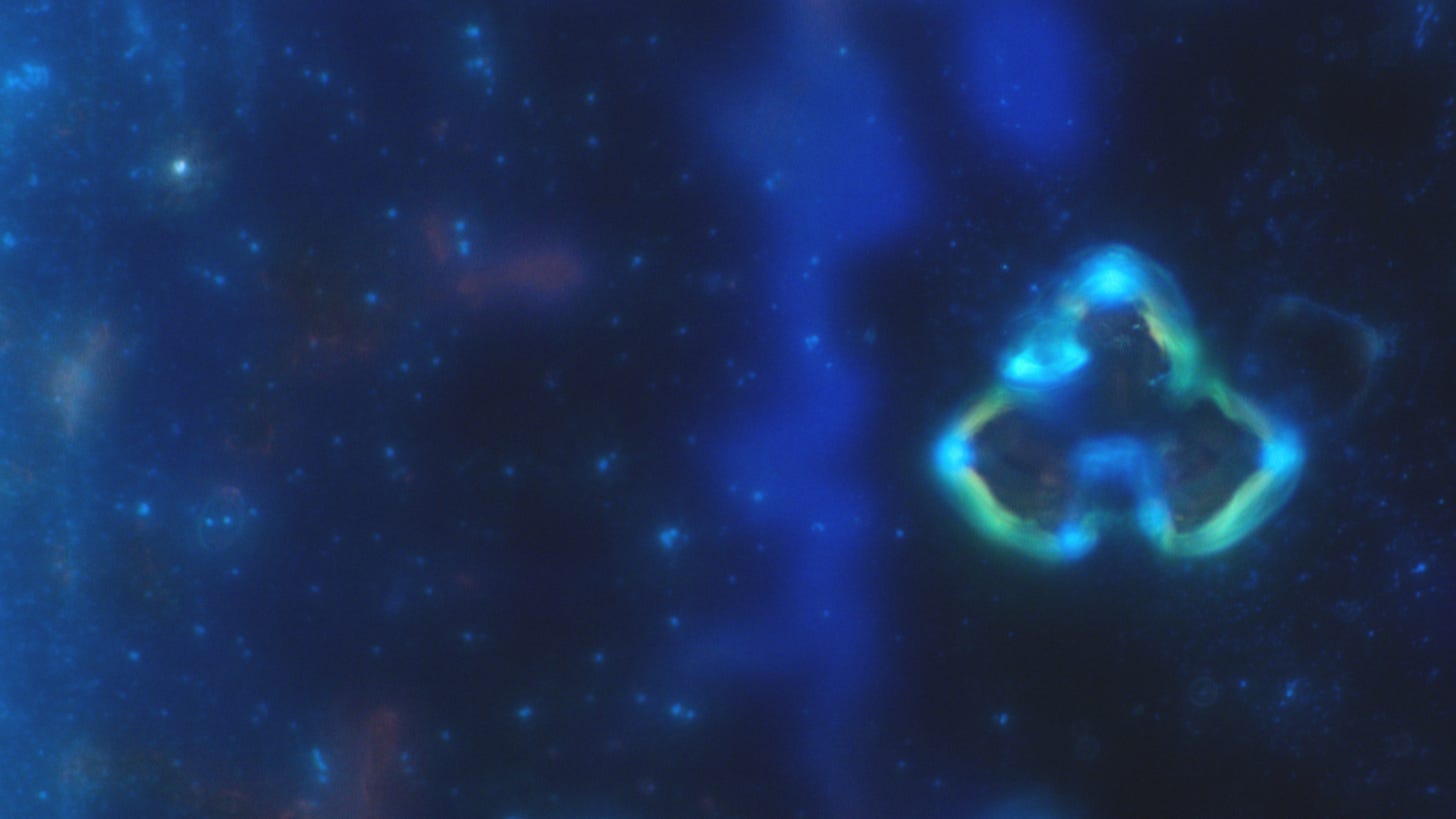
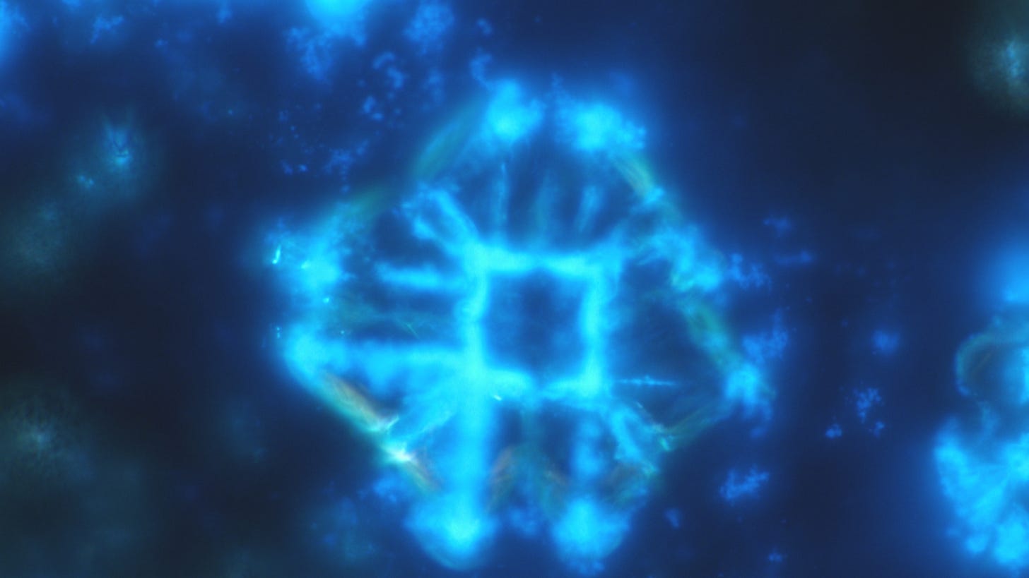
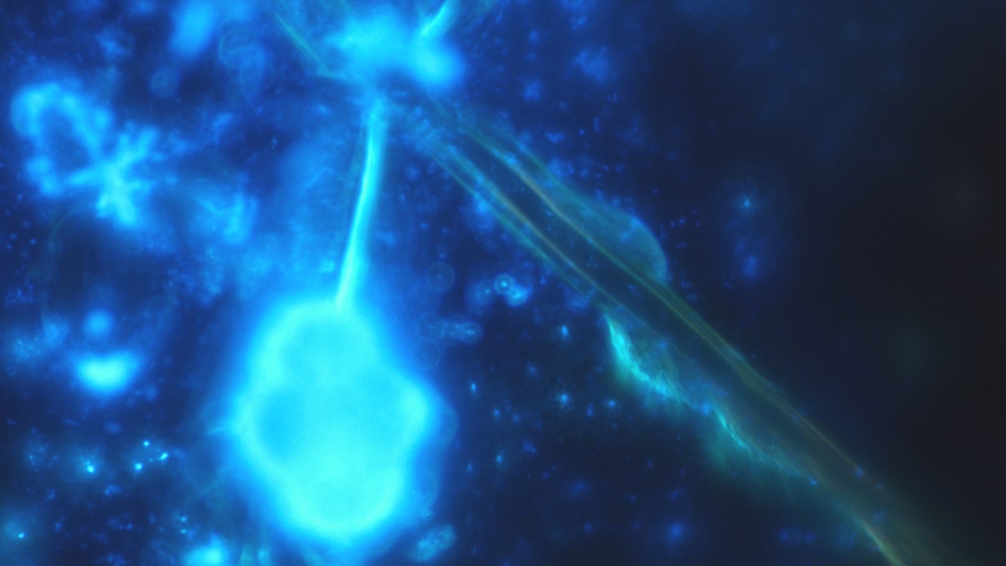
Standardized preparation of a urine sediment and direct examination under the dark-field microscope. 10 ml of fresh urine is centrifuged at 400 g for about 5-8 minutes.
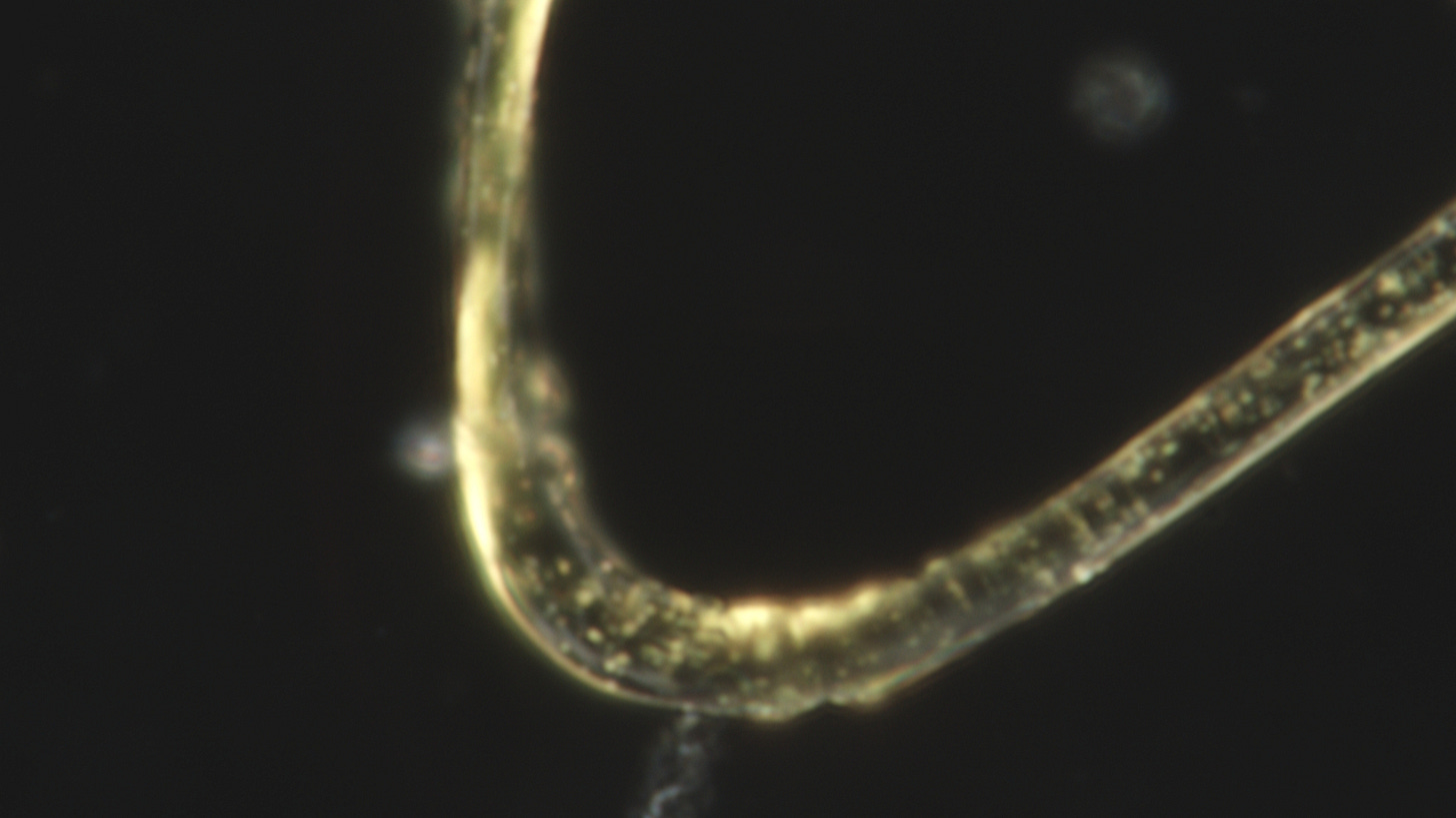
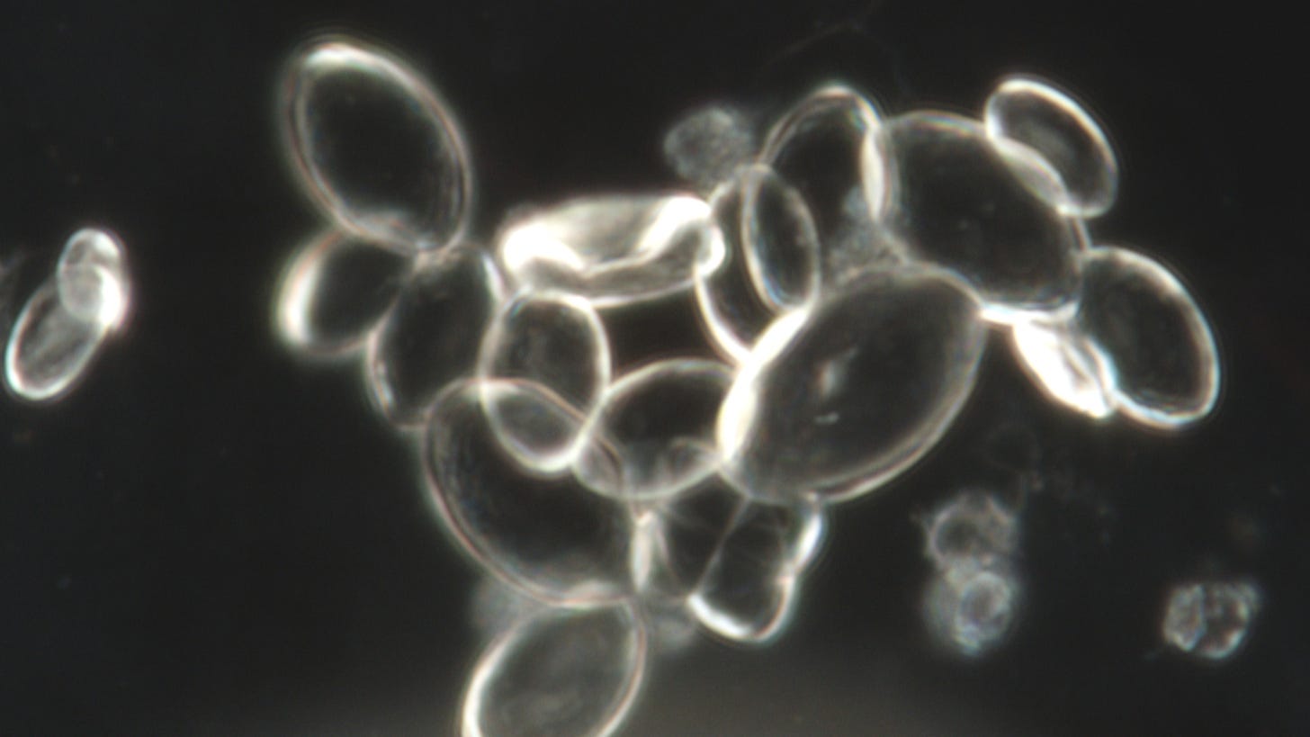
In summary, there are some thread-like structures and some fungal-like structures that also tend to occur together. In between, normal squamous epithelium.
Thread-like structures with yellow spots – at first I thought it had something to do with the urine. But later on, I came across a structure in the blood that was completely yellow:
These fungi are also not expected in fresh sediment.
An arrow construction... At the back, a tubular structure can clearly be seen, which has formed in the middle of the cells.
The sediment one day later in its crystallization:
A little urine sediment quiz:
You are welcome to book a subscription for the Remote Viewings directly at:
For your help and support here on Substack you will also get access to my Future Targets, Extra Targets and the first blueprint for the LaserCube on my website.
I have set up a Telegram channel - where I post new images from darkfield microscopy. If you would like to browse through new pictures in between, you are welcome to register.
Bitcoin QR-Code


